 The Musical Brain
- Resources from Eric Jensen
(The Brain Store - http://www.thebrainstore.com/store/
)
- Teaching with the Brain in
Mind and Music with the Brain in Mind - Eric Jensen
- Teaching With the Brain
in Mind balances theory and research with tips and techniques for
applying what we now know about the brain and learning to everyday
classroom practice. From its primer on brain biology to its in-depth
discussions of emotion, memory, and recall, this popular volume is
invaluable for educators looking to better reach students through truly
brain-compatible teaching.
- Music With the
Brain in Mind - Although compelling evidence supports the value of
the musical arts in school, many of us still fight for its inclusion.
This timely new resource translates the latest brain and music research
and provides practical strategies for incorporating the musical arts at
all levels.
- Read the Excerpt from Teaching
with the Brain in Mind (ASCD) - http://www.ascd.org/readingroom/books/jensen98book.html
Auditory - Learning through the Sense of
Hearing
How
does the brain process music?
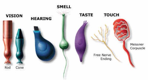
 The
Five Senses
The
Five Senses
How do we hear?
"Sound waves enter your ear canal and hit
your ear drum. This makes it vibrate. Three tiny bones in your middle ear link
the vibrating ear drum with the inner part of your ear. The last of these bones
is connected to a tiny bone structure that looks a bit like a snail shell, but
is about the size of a pea. It is called the cochlea (pronounced cock-lee-ah).
Your cochlea is filled with a liquid that carries the vibrations to thousands of
tiny hair cells. Each cell is tuned to a particular sound (or frequency). As
these little hair cells move in the fluid, they carry a message to the nerve
that is connected to your brain, which turns this signal into what you hear. All
this happens in a fraction of a second." Resource: http://www1.mydr.com.au/default.asp?article=3361
Seeing, Hearing, and Smelling
the World - Howard Hughes Medical Center
http://www.hhmi.org/senses/
This is a great website
presenting research on how we hear and how the brain processes sensory
input. These readings are optional, but they provide some wonderful
resources for understanding how we know the world through auditory
experiences. Here's a quote from "Sensing Change in the
Environment." "Everything we know
about the world comes to us through our senses.
Traditionally, we were thought to have just five of them—sight, hearing,
touch, smell, and taste. Scientists now recognize that we have several
additional kinds of sensations, such as pain, pressure, temperature, joint
position, muscle sense, and movement, but these are generally included under
"touch." (The brain areas involved are called the "somatosensory"
areas.)"
Brain Activity by
Age - Stages of Development Through Sensory Experiences in the First Year
|
|
Learning through the Senses -
Great Sites for Kids
How the Brain Responds to
Music Sound Waves
- Sound waves connect with hair
neurons that match the frequency.
- The frequency activates
neurons and mechanical energy is turned into electrical energy
- Neurons project to the
brainstem in the cochlear nucleus of the medulla where the location of sound
is determined.
- Projections from the auditory
cortex send information back to the cochlea to aid in auditory
discrimination
- Electrical package (music)
sent to thalamus
- These impulses flow to the
auditory cortex on the left side of the brain and light up the brain during
PET or MRI scans
- Brain has developed elaborate
neural networks that process the components of music - pitch, timbre,
harmony, and rhythm.
- Music is processed in the
brain - sensed, sorted, categorized, recognized, and responded to as quickly
as the neurons can fire, connect, and oscillate.
|
-
Our
non-dominant hemisphere processes harmonic structure, interval,
quality, timbre, and the spatial, temporal, and long-term patterns of
music. (right for most people)
-
Our
dominant hemisphere recognizes short term signatures, rapid variance
in volume, rapid and accurate pitch trajectory, pacing, and lyrics
(left for most people
|
Two Cerebral Hemispheres - Left and Right
- Connected by bundles of nerve fibers
- Allows each side of the brain to exchange information more freely
- Although each side processes things differently, the early concept of left
brain/right brain is outdated
- Left-handed and Right-handed
people use differing parts of the hemisphere for some activities
- Left Hemisphere
- Processes things more in parts and sequentially
- Musicians process music in left hemisphere
- Right Hemisphere
- Music and Arts have been considered right-brain "frills" but
trained musicians use more left-brain and novice musicians use more
right.
- Higher-level mathematicians, problem solvers, and chess players
actually have more right-brained activity, but beginners use more left
brain.
|
|
The Thalamus
|
The thalamus is often thought of as the individual consciousness - the
"You"
- Narrow bands across the top middle of the brain
- Sensory Cortex - Monitors skin receptors
- Motor Cortex - Needed for Movement
- Cerebellum
- Latin for "the little brain"
- Back lower area of the brain
- Responsible for balance, posture, motor movement, and some areas of
cognition
- Thought to include the essential long-term memory traces for motor
learning.
|
|
Broca |
Music is
processed differently for different people depending on kind of music and
musical background.
- Familiar music activates
Broca's area (left hemisphere)
- Rhythm notes are
activated in Broca's area and the cerebellum
- Harmony activates the
left side of the brain more than the right in the inferior temporal
cortex.
- Timbre activated the
right hemisphere (the only musical element that did)
- Pitch activated an area
on the left back of the brain - the precuneus.
- Melody activated both
sides of the brain.
- Composite listening -
Left and Right Hemisphere - Auditory Cortex
- Understanding lyrics -
Wernicke's Area
|
|
Wernicke's
Area - Lyrics |
Auditory
Cortex - Melodic Contour |
 Learning Changes the Brain!
- Some kind of stimulus to the
brain starts the learning process.
- The stimulus is sorted and
processed at several levels.
- Results in formation of
memory.
- Either doing something we
already know how to do - or we are doing something new.
- Stimulation is doing something
new - lighting up the brain scan.
- Once a task is learned, the
brain lights up less.
- Neurons (brain cells) make
connections between different parts of the brain.
- Information is carried inside
a neuron by electrical pulses and transmitted across the synaptic gap from
one neuron to another by chemicals called neurotransmitters.
- Learning is a critical
function of neurons.
- Dendritic branching helps make
connections between cells.
- As cells connect with other
cells, synapses occurs.
- New synapses appear after
learning.
- Repeating earlier learning
makes neural pathways more efficient through myelination (fatty substances
formed around axons)
| Brain
Activation with Different Stimulation and Levels of Activity
Performing music
makes neural connections between various parts of the brain.
Auditory and motor activities take place through playing an
instrument. Rhythm and melody instruments require motor
coordination. Reading music involves visual activity. Singing
songs and recalling or reading lyrics activate language processing areas
of the brain. Dancing or moving to the rhythm of music stimulates
the brain's motor areas.
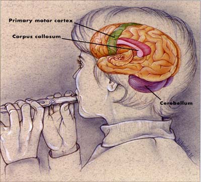
Brain research
provides support for emphasizing the visual and performing arts in the
classroom!
|
|
Auditory
Activity
|
Motor
Activity
|
|
Visual
Activity

|
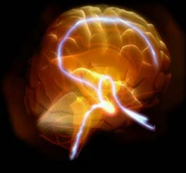
The Resting
Brain
|
| What
a Pet Scan Can Do
http://www.epub.org.br/cm/n01/pet/pet.htm
|
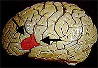
Language Processing
A good example of the fantastic
imaging capabilities of PET is shown in the images at the left, made by Dr. Marcus
Raichle, at the Neuroimaging Lab or
the Washington University School of Medicine, St Louis, USA.
These scans "were taken
under two different conditions. In the first one (uppermost image), an
individual was hearing a text, in order to learn a new language task. The color
map shows the regions of the brain which were activated by this task, in other
words, where there were cells working more than in their resting state, with a
higher metabolism (using more energy and more blood flow). The PET machine shows
the degree of activity in several tones of color, like in a rainbow. Yellow and
red regions are "hotter", that is, they indicate a higher cell
activity. Blue and black regions show decreased activity or none at all. While
obtaining this image, the patient was still unpracticed at the language learning
task. The highest brain activities are shown in an area called temporal lobe,
responsible for the hearing perception, and in another area called prefrontal
cortex, responsible for understanding language.
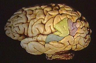
In the second condition
(lowermost image), the same individual has now learned the language task and is
spelling out. You can easily see in the color map that two different regions of
the brain were activated in each condition. Now the activity is concentrated in
the area of the cortex which is responsible for the motor control of voice, the
so-called area of Broca, so named because it was discovered by a French
physician named Paul Broca, in the turn of the century. Thus, the functional map
obtained with PET closely corresponds with what we know about the brain's
functional neuroanatomy, discovered by other methods. The difference here is
that we can actually obtain a real-time image of brain function."
|
We
now know much more about how the brain functions due to technological
advancements in "Neuroscience."

Explore these online
resources on the brain!
Neuroscience - Developing
through 1970's, 1980's, and 1990's
- Technology paved the way for
paradigm shift
- Enabled researchers to
understand and see inside the brain.
- Brain scanners developed -
Brain Imaging Technology
- Magnetic Resonance Imaging
(MRI)
- Positron Emission
Tomography (PET) - Radioactive glucose used to determine activity in
different parts of the brain
- International Society of
Neuroscience established - 1969
- Computerized Electrodes
- Electroencephalography
(EEG) - gives us readings about electrical output of the brain
- Detect brainwave patterns
- Can detect abnormal
cerebral functions (seizures or dementia)
- Can help us detect brain
activity during problem solving
- Autopsies
- Show weight, stages of
development, and amount of decay or lesions
- Dendritic activity shows
how brains physically changes with different tasks
- Spectrometers
- Measure specifics of brain
chemicals or neurotransmitters as activity occurs
- Levels of
neurotransmitters present in different brain lobes.
|
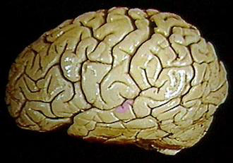
Facts
About the Human Brain
|
- Weighs approximately 3 pounds
- Mostly water - 78%
- Fat - 10%
- Protein - 8%
- Soft enough to cut with a butter knife
- Grapefruit-sized organ
- Outside of the brain
- Convolutions or folds
- Wrinkles are part of the cerebral cortex
- Folds allow maximum surface area
- Makes up critical portion of the nervous system
- Nerve cells connected by nearly 1 million miles of nerve fibers
- Has the largest area of uncommitted cortex of any species giving humans
flexibility for learning.
- Brain consumes about 20% of the body's energy
.
- The Brain uses about 1/5 of the body's
oxygen.
- The Brain gets about 8 gallons of blood each hour (supplying nutrients like glucose,
protein, trace elements, and oxygen).
- Brain needs 8-12 glasses of water a day for optimal functioning.
- Two kinds of brain cells:
- Glia - (Greek word meaning
glue)
- 90% of the brain
cells
- Less known about glia
cells
- No cell body
- Remove dead brain
cells and give structural support
- Neurons (Greek word
meaning bowstring)
- 100 billion neurons in
human brain
- Neurons essential to
performing the brain's work
- Consist of a compact
cell body, dendrites, and axons
|
Lobes - Four Areas of the Brain
Frontal Lobe
- Area around your forehead
- Involved in purposeful acts like judgment, creativity, problem solving,
and planning.
Parietal Lobe
- Top back area of the brain
- Processes higher sensory and language functions
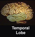
Temporal Lobe
- Left and right side above and around the ears
- Primarily responsible for hearing, memory, meaning, and language.
- Some overlap in functions of the lobes.
Occipital Lobe
- Back of the brain
- Primarily responsible for vision
Territory in the Middle of the Brain - The Emotional Area of the Brain
- 20% of the brain by volume
- Sometimes called "The Limbic System"
- Responsible for
- Emotions
- Sleep
- Attention
- Body regulation
- Hormones
- Sexuality
- Smell
- Responsible for production of most of the brain's chemicals
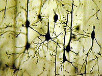
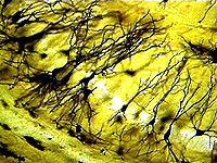
Gallery of Neurons - http://faculty.washington.edu/chudler/gall1.html
Dendrites and Axons
Neurons have specialized projections
called dendrites and axons. Dendrites bring information to the cell body
and axons take information away from the cell body. Information
from one neuron flows to another neuron across a synapse. The synapse is a small
gap separating neurons. (The Synapse - http://faculty.washington.edu/chudler/synapse.html)
- Dendrites
- Responsible for
information processing
- Fibers convert chemical
and electrical signals back and forth
- A normal functioning
neuron is continuously firing, integrating and generating information
- Hotbed of activity
- Many dendrites extend from
each neuron
- Each dendrite has one axon
that splits to subdivide itself and connect to other dendrites
- Information flows from
cell body down to axon to the synapse.
- Branch-like extensions
that grow outward when the environment is enriched.
- Neurotransmitters -
chemicals
- Axon
- Conducts information in
the form of electrical stimulation
- Transports chemical
substances
- The thicker the axon, the
faster it conducts electricity and information.
- Myelin (fatty substance)
forms around well-used axons ("myelinated")
A neuron typically has many
dendrites and one axon. The dendrites branch and terminate in the vicinity of
the cell body. In contrast, axons can extend to distant targets, more than a
meter away in some instances. Dendrites are rarely more than about a millimeter
long and often much shorter. Neurons communicate through specialized junctions
called synapses.
Axons typically make synapses with other neurons through specialized
enlargements near their terminals. These synapses can occur on the cell bodies
or the axons of other neurons, but most frequently they occur on dendrites.
Thus, the dendrites of a neuron provide a surface for receiving synaptic inputs
from other neurons. The arbor formed by the dendrites of a neuron often has a
characteristic shape as determined by the spatial domains into which the
dendrites family. (From Morphology of Dendrites - http://synapses.mcg.edu/anatomy/dendrite/dendrite.stm
- The Human Brain Project.)
Brain Activity Hotspots
See Additional Course
Documents for More Research Resources and Websites of Interest
Brain Websites
Music and the Brain Resources
Educational Articles from
Educational Leadership (ASCD)
Research on Child Development
- Society has historically
placed emphasis on the arts as a way to develop and stimulate the senses of
young children at various stages of development.
- Concepts of developmentally
appropriate practice with young children came out of the European scientific
research which resulted in "kindergarten" and early childhood
education.
- Human Intelligence Historical
Influences from University of Indiana - http://www.indiana.edu/%7Eintell/map.shtml

Copyright 2003
by Carla Piper, Ed. D.
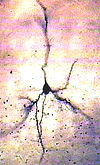 Neurons
- Brain Cells
Neurons
- Brain Cells